

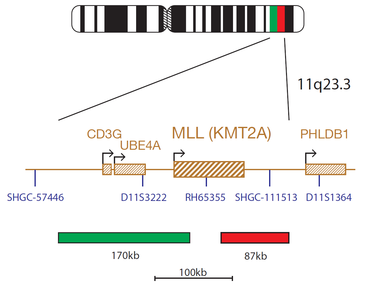
The KMT2A product consists of an 87kb probe, labelled in red, covering a region telomeric to the KMT2A gene including the marker SHGC-111513 and a green probe covering a 170kb region centromeric to the KMT2A gene spanning the CD3G and UBE4A genes.
Microscope image
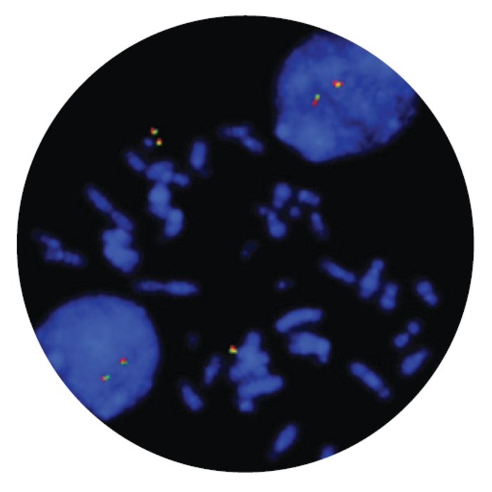
The CDA-LPH013 KMT2A Breakapart FISH Probe Kit PDx targets the KMT2A gene, with a probe design which flanks the KMT2A breakpoint region. This design detects the presence of a rearrangement involving the KMT2A gene, irrespective of the rearrangement partner; a positive breakapart signal pattern with CDA-LPH013 KMT2A Breakapart FISH Probe Kit PDx indicates the presence of a KMT2A rearrangement.
The KMT2A (lysine methyltransferase 2A) gene at 11q23.3 is commonly rearranged in acute leukemias, especially in infant leukemia. KMT2A rearrangements can be detected in approximately 70%-80% of infants with acute lymphoblastic leukemia (ALL) and in 5-10% of pediatric and adult ALLs1,2. They can also be found in more than 50% of infants with acute myeloid leukemia (AML) and is also seen in 10% of adolescents and 2-3% of adults with AML. KMT2A rearrangements are also seen in mixed-phenotype acute leukemia, a rare type of acute leukemia more common in infants and children than adults2. To date, more than 90 partners have been identified with the most common partner genes being AFF1 (4q21), MLLT3 (9p22) and MLLT1 (19p13.3)2.
The KMT2A gene encodes for a transcription factor which plays an important role in embryonic development and hematopoiesis as an important epigenetic regulator. The KMT2A rearrangements lead to the expression of KMT2A fusion proteins that interact with the nuclear protein menin. This KMT2A fusion protein – menin interaction leads to aberrant expression of HOX genes and MEIS1, causing a hematopoietic transformation block and leukemic transformation3,4,5.
The CDA-LPH013 KMT2A Breakapart FISH Probe Kit PDx is a fluorescence in situ hybridization (FISH) test used to detect rearrangement of the KMT2A region on chromosome 11 at location 11q23.3 in 3:1 methanol/glacial acetic acid fixed bone marrow specimens from patients with relapsed or refractory acute leukemia with KMT2A rearrangement.
The assay is indicated for detecting the presence of rearrangements involving the KMT2A region as an aid in identifying those patients for whom treatment with Revumenib is indicated. The CDA-LPH013 KMT2A Breakapart FISH Probe Kit PDx is not intended for monitoring of residual disease.
The test is for prescription use only.
|
Detection of |
KMT2A rearrangements |
|
Positive signal pattern |
1F1R1G |
|
Negative signal pattern |
2F |
|
Sample type |
3:1 methanol / glacial acetic acid fixed bone marrow specimens |
|
Patient age |
Any / not specified |
|
Patients with |
— Relapsed / Refractory — Acute myeloid leukemia (AML) — Acute lymphoblastic leukemia (ALL) — Mixed-phenotype acute leukemia (MPAL) |
|
Aid in identification of patients for whom treatment with [..] is indicated |
Revumenib |
This device is designed to detect rearrangements with breakpoints in the region bounded by the red and green clones in this probe set, which includes the KMT2A gene. Breakpoints outside of this region, or variant rearrangements wholly contained within this region, may not be detected with this device.
This device is not intended for monitoring of residual disease or for use as a stand-alone diagnostic test, prenatal test, population-based screening test, near-patient test, or self-test.
This device has not been validated for sample types, disease types, or purposes outside of those stated in the intended use.
Reporting and interpretation of FISH results should be performed by suitably qualified staff.
Reporting and interpretation of FISH results should be consistent with professional standards of practice and should take into consideration other clinical and diagnostic information. Failure to adhere to the protocol may affect the performance and lead to false results.
This device is intended for laboratory professional use only.
→ Download
Find certificate of analysis documentation for our CytoCell FISH probes
In our hands, CytoCell FISH probes have proven to be of the highest quality with bright, easy to interpret signals, thus providing confidence in our results. OGT's customer support is outstanding, as their staff are extremely knowledgeable and truly care about their customers and their customers’ needs.

Jennie Thurston
Director of Cytogenetics, Carolinas Pathology Group, USA
I first came across CytoCell FISH probes in a previous lab I worked in and I was struck by the quality of the products. Since this time, I have been recommending and introducing CytoCell probes across all application areas — now they are the primary FISH probes used in our lab. They have an excellent range of products and their ready-to-use reagent format saves considerable time.

Elizabeth Benner
Medical Technologist, University of Arizona Health Network, USA
Our lab has been using a wide range of CytoCell FISH probes for a number of years, and have been increasing this range all the time. The probes have clear bright signals and show good reproducibility. CytoCell provides fast delivery of catalog probes, and are very responsive when we have any queries or problems with their products.

Bridget Manasse
Addenbrookes Hospital, Cambridge University Hosiptals NHS Foundation Trust, UK
The quality and consistency of CytoCell’s probes means I can trust the results, and my clients get their results in a timely manner.

Dr. Theresa C. Brown
Director, Cytogenetics Laboratory, Hayward Genetics Center, Tulane University School of Medicine, USA
It was very important for us to have more consistent results with our probes — easy-to-read bright signals and a range of vial sizes, which is much more cost-effective.

Janet Cowan, PhD
Director of the Cytogenetics Laboratory, Tufts Medical Center, USA
Not only do CytoCell offer an extensive range of high-quality FISH probes, the customer support is also excellent — providing fast access to all the probes I need. The probes are highly consistent with bright signals allowing easy scoring of results.
Dr. Eric Crawford
Senior Director, Genetics Associates Inc., USA
We have been working with CytoCell fish probes for two decades because of their excellent clarity and intensity regardless of the size of the probe. It is so clear and simple to detect.
Dr. Marina Djurisic
Head of Laboratory of Medical Genetics, Mother and Child Health Care Institute of Serbia “Dr Vukan Cupic”, Belgrade, Serbia
The quality and reproducibility of results using the CytoCell kit has been vital in accurately detecting co-deletions in our glioma investigations. We now have a cost-effective test that we can rely on that is also easy to use and interpret. We've been consistently impressed with this kit - not to mention the support offered by OGT's customer service, and have completely transitioned over to CytoCell probes.
Gavin Cuthbert, FRCPath
Head of Cancer Cytogenetics, Northern Genetics Servce, Newcastle, UK
 Visit International site
 Visit Canada site