


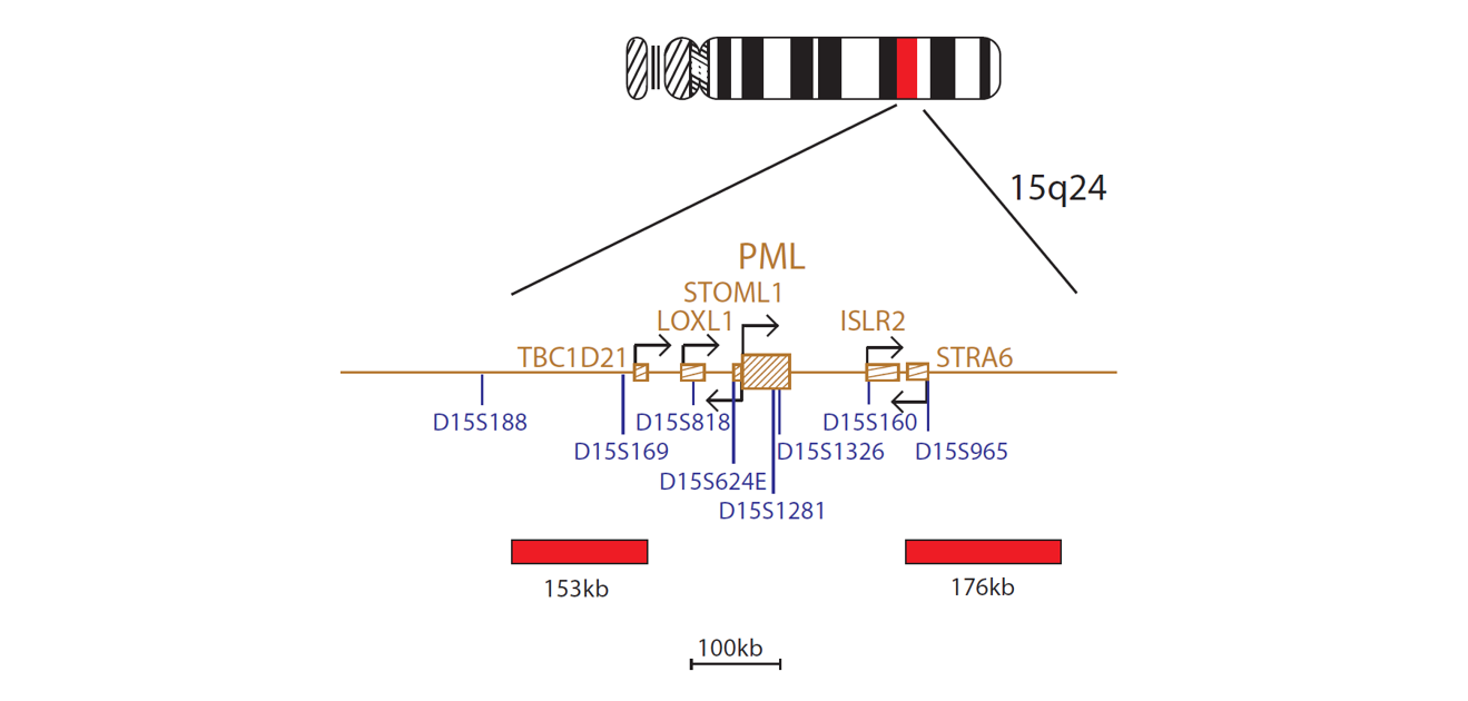
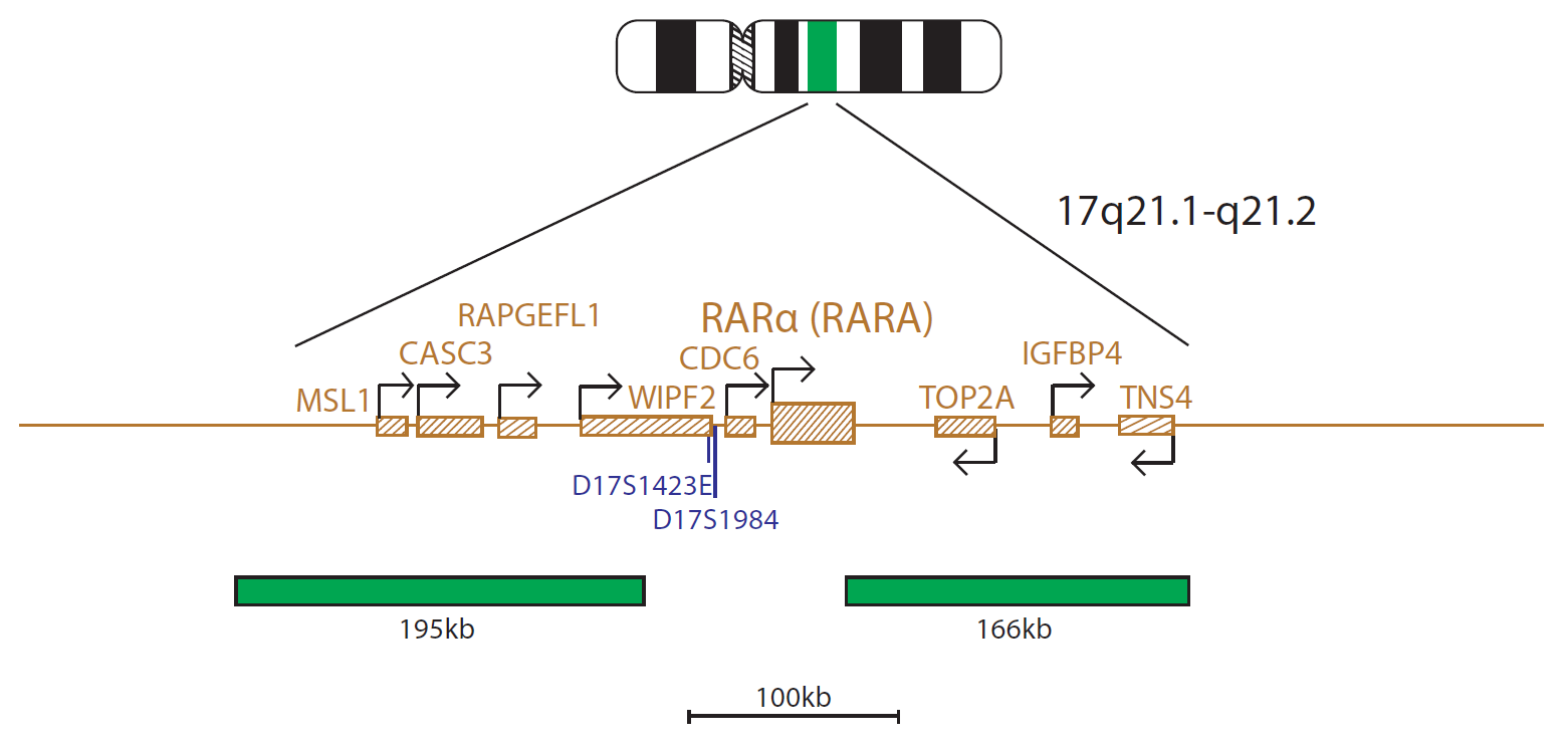
The PML probe mix, labelled in red, consists of a 153kb probe centromeric to the PML gene that covers the marker D15S169 and a 176kb probe telomeric to the PML gene that covers the marker D15S965. The RARα (RARA) probe mix, labelled in green, consists of a 195kb probe centromeric to the RARα (RARA) gene that spans the CASC3 gene and a 166kb probe that covers the telomeric end of the RARα (RARA) gene as well as the TOP2A, IGFBP4 and TNS4 genes.
Microscope image
The PML (promyelocytic leukaemia) gene is located at 15q24.1 and the RARA (retinoic acid receptor alpha) gene is located at 17q21.2. The translocation t(15;17)(q24;q21) gives rise to the PML::RARA fusion gene and is the diagnostic hallmark of acute promyelocytic leukaemia (APL).
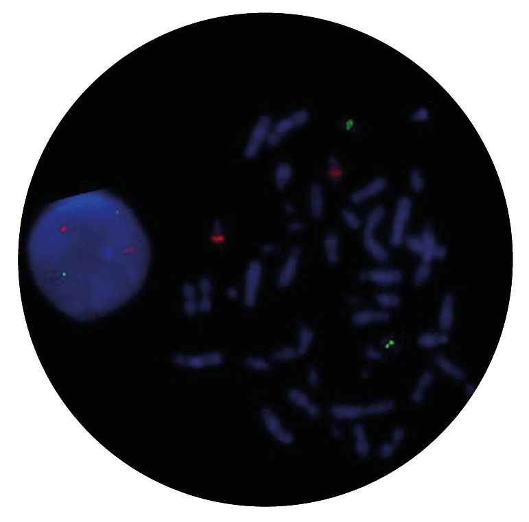
This FAST PML/RARα FISH probe allows rapid detection of the rearrangement, with only one hour of hybridisation required.
The PML::RARA fusion gene is created by the t(15;17)(q24;q21) translocation, found in more than 90% of cases of APL, a leukaemia that comprises 5-8% of cases of acute myeloid leukaemia (AML)1,2. In a subset of cases, variant RARA translocations can be observed. Known fusion partners include NPM1 at 5q35, NUMA1 at 11q13, ZBTB16 (PLZF) at 11q23, STAT5B at 17q21, PRKAR1A at 17q24, FIP1L1 at 4q12 and BCOR at Xp113,4,5.
PML and RARA have both been implicated in normal haematopoiesis. PML possesses growth suppressor and proapoptotic activity whereas RARA is a transcription factor that mediates the effect of retinoic acid at specific response elements6. PML::RARA fusion protein behaves as an altered retinoic acid receptor with an ability of transmitting oncogenic signaling7.
Immediate treatment of APL patients is critical, due to fatal coagulation disorders and life-threatening haemorrhage in diagnosis. Prior to the introduction of all-trans-retinoic-acid (ATRA) and arsenic trioxide (ATO) in APL treatment protocols, the disease had a poor prognosis; however, since the introduction of these therapies, the overall survival rate has improved dramatically, with nearly 90%5 of patients cured. Patients with variant RARA translocations show variable sensitivity to treatment, with some patients showing resistance to treatment protocols3,5. It is therefore important to differentiate between APL patients with PML::RARA fusion and those patients with variant RARA translocations.
The CytoCell® FAST PML/RARα (RARA) Translocation, Dual Fusion Probe is a qualitative, non-automated, fluorescence in situ hybridisation (FISH) test used to detect chromosomal rearrangements between the 15q24 region on chromosome 15 and the 17q21.1-q21.2 region on chromosome 17 in Carnoy’s solution (3:1 methanol/acetic acid) fixed haematologically-derived cell suspensions from patients with confirmed or suspected acute myeloid leukaemia (AML).
This device is designed as an adjunct to other clinical and histopathological tests in recognised diagnostic and clinical care pathways, where knowledge of PML::RARA translocation status would be important for clinical management.
This device is designed to detect rearrangements with breakpoints in the region covered by the red and green clones in this probe set, which includes the PML and RARA regions. Breakpoints outside this region, or variant rearrangements wholly contained within this region, may not be detected with this device.
This device is not intended for: use as a stand-alone diagnostic, use as a companion diagnostic, prenatal testing, population-based screening, near-patient testing, or self-testing.
This device has not been validated for sample types, disease types, or purposes outside of those stated in the intended purpose.
It is intended as an adjunct to other diagnostic laboratory tests and therapeutic action should not be initiated on the basis of the FISH result alone.
Reporting and interpretation of FISH results should be performed by suitably qualified staff, consistent with professional standards of practice, and should take into consideration other relevant test results, clinical and diagnostic information.
This device is intended for laboratory professional use only.
Failure to adhere to the protocol may affect the performance and lead to false positive/negative results.
Find certificate of analysis documentation for our CytoCell FISH probes

Our lab has been using a wide range of CytoCell FISH probes for a number of years, and have been increasing this range all the time. The probes have clear bright signals and show good reproducibility. CytoCell provides fast delivery of catalogue probes, and are very responsive when we have any queries or problems with their products.

Bridget Manasse
Addenbrookes Hospital, Cambridge University Hosiptals NHS Foundation Trust, UK
In our hands, CytoCell FISH probes have proven to be of the highest quality with bright, easy to interpret signals, thus providing confidence in our results. OGT's customer support is outstanding, as their staff are extremely knowledgeable and truly care about their customers and their customers’ needs.

Jennie Thurston
Director of Cytogenetics, Carolinas Pathology Group, USA
I first came across CytoCell FISH probes in a previous lab I worked in and I was struck by the quality of the products. Since this time, I have been recommending and introducing CytoCell probes across all application areas — now they are the primary FISH probes used in our lab. They have an excellent range of products and their ready-to-use reagent format saves considerable time.

Elizabeth Benner
Medical Technologist, University of Arizona Health Network, USA
We have been working with CytoCell fish probes for two decades because of their excellent clarity and intensity regardless of the size of the probe. It is so clear and simple to detect.
Dr. Marina Djurisic
Head of Laboratory of Medical Genetics, Mother and Child Health Care Institute of Serbia “Dr Vukan Cupic”, Serbia
The quality and consistency of CytoCell’s probes means I can trust the results, and my clients get their results in a timely manner.

Dr. Theresa C. Brown
Director, Cytogenetics Laboratory, Hayward Genetics Center, Tulane University School of Medicine, USA
It was very important for us to have more consistent results with our probes — easy-to-read bright signals and a range of vial sizes, which is much more cost-effective.

Janet Cowan, PhD
Director of the Cytogenetics Laboratory, Tufts Medical Center, USA
Not only do CytoCell offer an extensive range of high-quality FISH probes, the customer support is also excellent — providing fast access to all the probes I need. The probes are highly consistent with bright signals allowing easy scoring of results.
Dr. Eric Crawford
Senior Director, Genetics Associates Inc., USA
The quality and reproducibility of results using the CytoCell kit has been vital in accurately detecting co-deletions in our glioma investigations. We now have a cost-effective test that we can rely on that is also easy to use and interpret. We've been consistently impressed with this kit - not to mention the support offered by OGT's customer service, and have completely transitioned over to CytoCell probes.
Gavin Cuthbert, FRCPath
Head of Cancer Cytogenetics, Northern Genetics Servce, Newcastle, UK
 Visit USA site
 Visit Canada site


